Some basics of Anatomy:
The aorta is the main artery in the human body, she was born in the output of the heart and descends into the thorax, and the abdomen to give many branches for different bodies.
It is divided into several portions:
The ascending aorta: giving the arteries to the heart: coronary arteries.
Aortic Lacrosse: gives the arteries to destinations of the 2 arms (sous-clavieres arteries) and the brain (carotid arteries and vertebral).
The descending thoracic aorta which gives many branches to the vertebrae and the spinal cord.
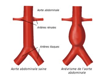
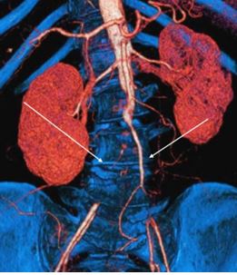
The abdominal aorta that gives the arteries to the viscera (trunks celiac, mesenteric arteries, upper and lower) and the renal arteries. It bifurcates to the navel to give the two iliac arteries at destination of the lower limbs.

The pathologies that can touch the aorta:
Aneurysms: the aorta may by place dilate: one speaks of aneurysm when she made more than 30 mm or once and a half its usual diameter.
There are many causes of aneurysm: mainly hypertension, tobacco, causing a weakening of the wall. Some family illness (Marfan disease) also promote his aneurysms.
The main risk of the aneurysm is that as they grow, they break causing an array of internal bleeding quickly, leading in the absence of support, to death.
These aneurysms are classified according to their location:
Aneurysm of the ascending aorta, aneurysm of the aortic Lacrosse, aneurysm of the thoracic aorta, best abdominal aneurysm, aneurysm of the aorta under kidney (the most common).
These aneurysms have a tendency to always grow. Furthermore more the bigger they are, the higher the risk that they break. And more they are big, their faster growth.
For a vein, a 50 mm of the abdominal aorta aneurysm risk of rupture of 5 to 10% per year for a growth of 5mm per year. A 70mm aneurysm presents an annual rupture risk by 30 to 40%.
Aortic dissection: the wall of the aorta is made up of several concentric layers (intima, media, weed). Sometimes, the two innermost layers (intima and media) may tear the outermost layer (adventitious) causing a dissection. The blood rushes in this tear causing the constitution of two channels of circulation: a true channel (the original light) and a fake tear from channel.
The consequences at the level of the thoracic aorta are multiple:
The tear causes a weakening of the arterial wall that may rupture.
When the fake channel extends all along the thoracic aorta and abdominal, dissection can compromise the circulation of all of the arteries which result from the aorta causing multiple complications (so-called malperfusion): paralysis of the 2 legs (the spinal cord ischemia), necrosis of the viscera (mesenteric ischemia), failure acute kidney, ischemia of the lower limbs.
Thrombosis: The presence of atheroma in aorta, favored by risk factors cardiovascular (smoking, high blood pressure,…), can cause stenosis. In general, this stenosis affects the aorta under kidney and exceptionally aorta upstream.
Eventually, this stenosis can gradually (over several years) become soiled and then completely clog causing aortic thrombosis.
Resulting symptoms pain walking or resting on both lower limbs (intermittent claudication). The disorders are possible. When they are associated with a limp called Leriche syndrome.
The evolution of the thrombosis is done by a progressive worsening of the perimeter of the market to cause pain permanent rest and term a gangrene in the Member table.
To make more clear exposition to deal with each of the major pathologies of independently:
- Aneurysm of the abdominal aorta
- Aneurysm of the thoracic aorta
- Thrombosis of the abdominal aorta, also known as Leriche syndrome
- Dissection of the descending thoracic aorta